Do polar microalgae get sunburned?

“Algal foam” made up by the haptophyte genus Phaeocystis. Cell size 3–6 μm. Photo: Angela Wulff
Aims
The aims of the suggested project are to study (I) the content of UV-absorbing compounds (mycosporine-like amino acids, MAAs) in phytoplankton along depth gradients (i.e. light gradients), (II) the relation between MAAs and typical light shielding pigments such as beta-carotene and diatoxanthin and (III) the accuracy of using biomarker pigments to describe the spatial distribution of different phytoplankton taxonomical groups.
Background
Phytoplankton are important microalgal primary producers in the coastal zone as well as in the open ocean. Algae at the surface are exposed to high irradiances of both photosynthetic available radiation (PAR, 400–700 nm) and ultraviolet-B radiation (UVBR, 280–320 nm), requiring strategies against photodamage. Such strategies involve the production of both photoprotective pigments, such as beta-carotene and diatoxanthin, as well as UV-absorbing compounds, e.g. MAAs (Karentz et al, 1991).
Recent trends in ozone depletion caused by atmospheric pollution most likely occur on much shorter time scales than the “natural” transitions that have been documented during the evolution of the earth’s atmosphere. Nowadays, an increased UVBR caused by ozone depletion is a well-known fact and is a threat at Nordic latitudes. In the Arctic part of Canada, a 45% thinning of the ozone layer was measured during spring 1997 (according to the Canadian Department of Environment). During the origin of life on earth no protective ozone layer had yet developed, and protection from harmful UVR should have been an important feature in the early evolution and natural selection of eukaryotic organisms. While terrestrial plants have developed more elaborate protection mechanisms (e.g. leaf shape and orientation), microalgae rely, more or less exclusively, on intracellular UV protection, such as the production of MAAs.
The deleterious effects of UVBR on marine microalgae are well recognised (e.g. Davidson et al., 1996, Wulff et al., 1999, Wängberg et al., 2001). Since microalgal species differ widely in tolerance, or capacity to adapt, to UVBR, it could even nowadays constitute a selective pressure, thereby changing the community species composition (Davidson et al., 1996). Furthermore, microalgae differ in their strategies for reducing UV exposure and limiting the amount of photodamage, including production of MAAs. MAAs are water soluble compounds with different UV absorption maxima between 310–360 nm and have been identified in most microalgal groups (Jeffrey et al., 1999). Microalgae have been shown to contain several different MAAs, and it is assumed that MAAs provide protection from UVR, analogous to the melanin pigmentation response of human skin.

Microalgal cells belonging to the haptophyte genus Phaeocystis. Photo: Pauline Snoeijs and Mats Kuylenstierna
In nature, most studies regarding MAAs have been performed in Antarctic waters, and so far no or very few studies have been conducted on Nordic microalgal species. In Antarctic waters it has been shown that phytoplankton within the upper mixed layer have higher concentrations of UV-absorbing compounds than phytoplankton found below the upper mixed layer (Vernet et al., 1994).
Phytoplankton identification and quantification is usually achieved through microscopic examination, which is very time-consuming and requires a high level of taxonomic skill. Furthermore, small cells, especially flagellates, belonging to nano- or picophytoplankton are easily overlooked or sometimes bonded together as one group. As an alternative to microscopic examination, photosynthetic pigments can be used for studying the composition and physiological status of phytoplankton, as certain pigments serve as taxon-specific indicators of major taxonomic groups. High performance liquid chromatography (HPLC) can be used to separate all relevant pigments, and is now a common tool, extensively used for estimating pigment concentrations in microalgal communities. In order to determine algal class abundance from group-specific pigments Mackey et al., (1996) developed the matrix factorisation program CHEMTAX. The program uses the steepest-descent algorithm to determine the best fit based on an estimate of pigment/chl a ratios for different algal classes.
The work on board
UVBR and PAR were measured in air 1–3 times a day using a photometer (International Light 1400A) with cosine corrected sensors. When possible the underwater light climate (downwelling irradiance) was measured with an underwater radiometer (Biospherical Instruments PUV-500). This instrument measured radiation at four discrete wavelength bands, centered at 305, 320, 340 and 380 nm, as well as the total amount of PAR (400–700 nm). The instrument was lowered from the aft end of the ship to a maximum depth of 60 m.
At 60 different stations, water samples were taken from the CTD rosette sampler or from GoFlo bottles. Generally, seawater was sampled from the surface (approx. 1–3 m depth depending on weather conditions) and from maximum chlorophyll fluorescence depth (chl max). If chl max was not possible to determine, seawater was sampled from 20 m depth. The chl max depth was determined using a handheld “mini CTD” equipped with a fluorometer. The water was filtered onto GF/F filters within 10–45 min after sampling, the filters were frozen in liquid nitrogen (–196°C) and transferred to a low temperature freezer (–80°C). Filtration took place in a dark, cool container to minimize the influence of light and temperature on the phytoplankton filtered. The filters are now being analysed for content and composition of photosynthetic pigments and MAAs. A total of approx. 600 water samples of 100–3 000 ml were filtered.

Positions of the different stations presented in figure 2.
In addition, approx. 120 samples were taken for onboard analyses of phytoplankton species composition (light microscopy). Usually this is not done onboard due to difficulties with vibrations from the ship and rough sea. Fixed samples often give a distorted picture of the species composition as several species get destroyed and the fixative makes it impossible to estimate the cell conditions. However, a more detailed identification will be performed after the cruise, therefore the phytoplankton were preserved with basic and/or acid Lugol’s solution.
Furthermore, the phytoplankton community composition will be analysed both through the use of biomarker pigments in CHEMTAX (see above) and through extensive microscopical analyses in order to compare the two different methods.
At 36 stations, phytoplankton primary productivity was measured (C-14-technique) in triplicate, with and without UVBR. One set of incubators was covered with Mylar foil to exclude UVBR and all samples were covered with cellulose diacetate film to exclude UVCR. The samples were incubated for 4 hours in the temperature controlled dark container under artificial light. The artificial light was adjusted to mimic the natural ratio between PAR and UVBR. The so called “blanks”, i.e. samples used to correct for uptake of 14C in the dark, were analysed on a scintillation counter onboard and were reanalysed together with the remaining samples after the cruise.
Preliminary results
The UVBR reached high intensities but the absolute values should be treated with care as the sensor is currently being calibrated. Generally, PAR penetrated down to the maximum depth measured (60 m) and the UVBR (320 nm) reached below 10 m depth.

Light measurements on deck measured daily around noon from 1 May to 3 June.
Samples for photosynthetic pigments and MAAs are currently under analysis and no results can be reported.
The onboard examination of living phytoplankton cells showed that the genus Phaeocystis (photo 1) dominated at several stations, particularly at stations 80–82 and 95 (for positions see table 1). This genus has been shown to kill fish due to mucus covering the colonies and getting stuck in the gills. Phaeocystis has earlier been shown to produce MAAs. Interestingly, we found diatom species that had earlier only been reported from the northern part of the Pacific. Very preliminary results from the more extensive species analyses show that stations 10, 11 and 14 were totally dominated by small flagellates in the size range 3–6 μm, so called nanoplankton. According to species diversity, diatoms dominated at these three stations.

Phytoplankton primary productivity as disintegrations/minute (DPM) from the water surface and chlorophyll fluorescence maximum depth (chl max). Treatments were –UVB (PAR+UVA) and +UVB (PAR+UVA+UVB). The data shown are from the 13 stations with highest primary productivity.
NB: Values from station 95 are out of range and should read 35 000/40 000 and 54 000/41 000 for the different treatments and surface and chl max, respectively.
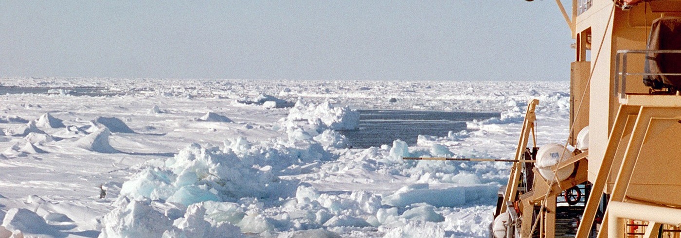
In general the primary productivitywas very low, with no statistically significant differences (ANOVA) between phytoplankton exposed to or protected from UVBR. This indicates that the phytoplankton are well adapted to UVBR. The highest primary productivity was associated with the ice edge, or regions where the ice had broken up.
Dates
1 May–30 May 2002
Participants
Principal investigator
Angela Wulff
Department of Marine Ecology, University of Gothenburg
Sweden
Anna Engelsen
Department of Marine Ecology, University of Gothenburg
Sweden
Mats Kuylenstierna
Department of Marine Ecology, University of Gothenburg
Sweden
Sten-Åke Wängberg*
Botanical Institute, University of Gothenburg
Sweden
*Not participating in the expedition
References
Davidson, A.T., Marchant, H.J. and de la Mare, W.K. 1996. Natural UVB exposure changes the species composition of Antarctic phytoplankton in mixed culture. Aquat. Microb. Ecol. 10, 299–305.
Jeffrey, S.W., MacTavish, H.S., Dunlap, W.C., Vesk, M. and Groenewoud, K. 1999. Occurrence of UVA- and UVB-absorbing compounds in 152 species (206 strains) of marine microalgae. Mar. Ecol. Prog. Ser. 189, 35–51.
Karentz, D. McEuen, F.S., Land, M.C. and Dunlap, W.C. 1991. Survey of mycosporine-like amino acid compounds in Antarctic marine organisms: potential protection from ultraviolet exposure. Mar. Biol. 108, 157–166.
Mackey, M.D., Mackey, D.J., Higgins, H.W. and Wright, S.W. 1996. CHEMTAX – a program for estimating class abundances from chemical markers: application to HPLC measurements of phytoplankton. Mar. Ecol. Prog. Ser. 144, 265–283.
Wängberg, S.Å., Wulff, A., Nilsson, C. and Stagell, U. 2001. Impact of UV-B radiation on microalgae and bacteria: a mesocosm study with computer modulated UV-B radiation addition. Aquat. Microb. Ecol. 25, 75–96.
Vernet, M., Brody, E.A., Holm-Hansen, O. and Mitchell, B.G. 1994. The response of Antarctic phytoplankton to ultraviolet radiation: absorption, photosynthesis, and taxonomic composition. Ant. Res. Ser. 62, 143–158.
Wulff, A., Nilsson, C., Sundbäck, K., Wängberg, S.Å. and Odmark, S. 1999. UV radiation effects on microbenthos – a four month field experiment. Aquat. Microb. Ecol. 19, 269–278.